Wednesday, April 27, 2016

Winter Work-study at SA (2015-2016): Adventures in Legacy Collections & 3D Scanning


Western work-study student, Kylie Kelly, has spent a couple of years preparing collections from the Museum of Ontario Archaeology for long term storage at SA. Today, her guest blog discusses her experience with one of SA's newly acquired legacy collections and some of the 3D scanning she has been learning with artifacts from that collection.

Author biography

Kylie Kelly is a work study student at the Museum of Ontario Archaeology and Sustainable Archaeology: Western. She is currently completing the last few weeks of her Bachelor’s degree in Classical and Medieval Studies at Western University. She is set to begin her Masters of Art History in the fall of 2016. Kylie became interested in working at SA during her time as an assistant curator at the Museum of Ontario Archaeology. She has been working simultaneously at both institutions since September of 2015.

Introduction

In the summer of 2015 Sustainable Archaeology received a legacy collection that contained tens-of-thousands of artifacts from nearly every period of Canada’s archaeological past-- a private collection donated to SA by Dr. Brian Deller and family. The collection represents the life’s work, and passion of Merle Franklin.  Mr.Franklin, a mostly self-taught archaeologist, focused much of his research on the archaeology of the lower Grand River. He carefully documented the objects as well as the spots on which they were found. Following the acquisition of the legacy collection, SA’s work study students, including myself, were tasked with reading through the pages of documentation, and sorting through the corresponding objects in the collection.
Slowly, the hand-made artifact trays and boxes started to fill the shelves at SA. On Monday I could be working on a box that was full of historic glass trade beads, and the next day I could be working with ancient lithics. The expansive collection contains everything from Paleo-Indian projectile points, to historic railroad ties-- it truly has it all.  A few weeks ago I went to retrieve another box to sort through, and two artifacts caught my attention. The artifacts, a fork and a knife, were like nothing I had seen before (see figures 1 and 2, below). The fork had thin and stubby prongs, similar to classical depictions of Poseidon’s trident. The knife was equally as fascinating, it had an elongated ivory handle with multiple metal rivets. I had just started training with our white light scanners under the tutelage of Nelson Multari, SA’s 3D Technician, and Hillary Kiazyk, a part-time staff member at SA and student in Western’s Anthropology Department. I thought that the cutlery’s interesting shapes, reflective surfaces, and straight edges would be a great challenge for me to capture accurately.

Object History

As I began scanning the fork/knife duo I became more interested in their history, and how they had ended up in a collection in Southern Ontario. The knife had a clear makers mark stamped onto it that read “Joseph Rodgers E. Sons, Cutlers to Her Majesty” (Figure 2). Joseph Rodgers & Sons Cutlery Company was started in 1764 in Sheffield, England. It appears that the company began by making elaborate pocket knives; and that by 1821 they gained enough attention that King George IV appointed them “Cutlers of the Royal Family.”[i]   The company’s favour continued over the span of five successive monarchs: King George IV, William IV, Queen Victoria, Edward II, and George V. I discovered that the donated set was made between 1837 and 1901, during the Reign of Queen Victoria. One of the most famous knives created by the company was the Norfolk Knife; it was produced during the same time period as the set at SA. But unlike the rest of the artifacts in the donated collection, the context of this set of cutlery – where it originated and how it ended up in this collection -- is regretfully unknown, as these particular items were donated un-provenienced.

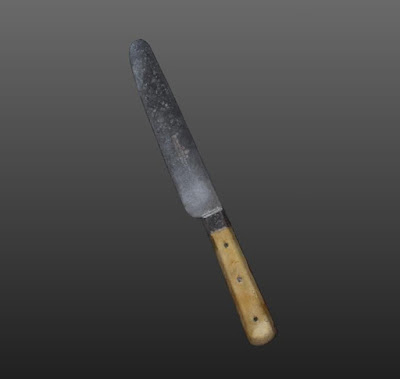
Figure 1: Knife and fork found in the Franklin/Deller collection.

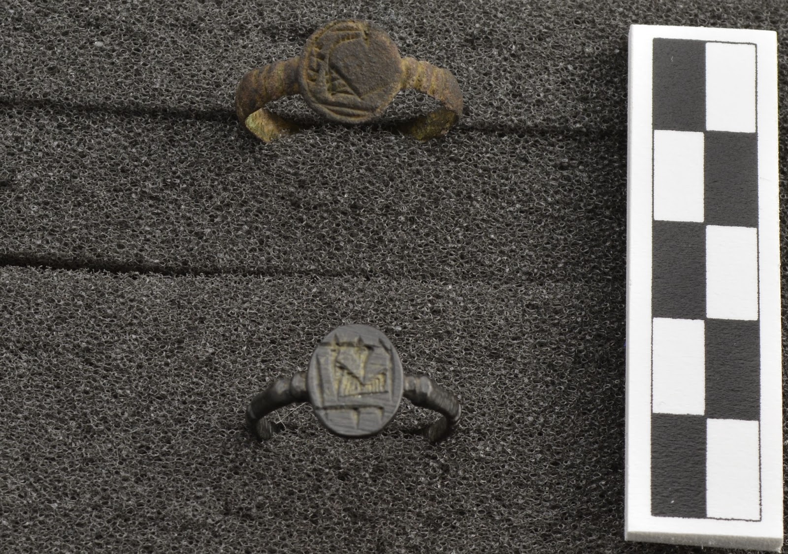
Figure 2: Maker's mark found on knife in Franklin/Deller collection.

Scanning Procedures

The 3D3 Systems (now LMI Technologies) structured light scanners at SA work by projecting a pattern of light onto the object. It then uses a rotating stage and cameras to capture the geometry of the projected lines in order to determine the shape and size of the surface of the object. An additional DSLR camera works to capture the texture, or color, of the artifact. The scanners are operated through Flexscan 3D 3.3 capture software, which allows the user to control the cameras, turntable, projector and model (for a detailed description of the white light scanning process at SA, see Ahmed et al. 2014). The nature of structured light scanning makes it difficult to capture reflective surfaces, as light projected onto the surface just bounces back off, leaving behind a fragmentary mesh, a problem I encountered repeatedly. Capturing a sharp edge is also challenging, as it requires scanning a mesh while utilizing lower cleanup settings in order to increase the amount of detail on the thin surfaces, thus there can be a lot of extra ‘noise’ captured in a scan that needs to be removed. To control the shine on the blade, I sprayed it with a non-corrosive powder to allow the white light to capture the surface, without bouncing off.  Due to the high learning curve associated with this technology, and this being my first independent scanning project, completing the preliminary scans of the knife took roughly 6 hours, and over 200 captures.  Finally, I was able to export the scan as a polygon (.ply) file to Geomagic Studio 2012 reconstruction software to refine the mesh and fill in any holes. I utilized a feature called mesh doctor that examines a 3D scan to detect and correct errors in mesh [ii].  Using this tool can result in a model that is “water tight”, meaning no holes are in the mesh. This water tight file is what we want to create if we are interested in producing a 3D print of a 3D scanned object.

http://rc.db.sa.uwo.ca/BloomCE/Knife/

The Take Away

As I begin my Master’s degree in Art History next fall, I am constantly amazed with how relevant new technologies, like 3D scanning, are across numerous fields of study. Part of my research as an Art Historian looks at how artwork was constructed; by using 3D imaging I developed a new understanding of the craftsmanship and time it took to complete something that looks deceivingly simple.  After the scan was complete I noticed many quirks in the artifact that I would have otherwise missed with the naked eye. The edge of the blade increases in size by a fraction of a millimeter the closer you get to the base; the surface of the blade has a distinct pattern of pitting on only one side; the handle’s rust deposits run parallel to the small fractures in the ivory. It is fascinating to me, given its broader context that this special set of monarch-endorsed cutlery ended up in an archaeology collection in southern Ontario – and it makes me wonder more about who owned it, and why and how it came to be included in the Franklin/Deller Collection—the possibilities of discovery are endless at Sustainable Archaeology!

Footnotes

 i "The History of Joseph Rodgers." - Egginton Bros Ltd. Accessed April 20, 2016. http://www.eggintongroup.co.uk/history/the-history-of-joseph-rodgers.html.
ii  For a detailed description of Mesh Doctor please visit: http://support1.geomagic.com/link/portal/5605/5668/Article/1012/What-is-the-Mesh-Doctor

References

"The History of Joseph Rodgers." - Egginton Bros Ltd. Accessed April 20, 2016. http://www.eggintongroup.co.uk/history/the-history-of-joseph-rodgers.html.

"The Sheffield Cutlery Map." Joseph Rodgers & Sons. Accessed April 20, 2016. http://www.sheffieldcutlerymap.org.uk/location/joseph-rogers-and-sons/.

2014       Ahmed, N., M. Carter and N. Ferris. Sustainable archaeology through progressive assembly 3D digitization. World Archaeology 46(1): 137-154.

Tuesday, March 1, 2016

Guest blog: New ways archaeologists ‘dig out’ answers


By: Victoria Nagy, MSc


After spending a field season conducting consultant archaeological field work in Ontario, I wanted to work in a research facility, an environment I enjoyed during my Masters studies. Therefore, I was excited to join the staff at Sustainable Archaeology: Western University. During my past undergraduate and graduate studies I had been exposed to digital archaeology and database development but I had never been able to directly apply those skills.

I was fascinated by the shear potential for research at Sustainable Archaeology since I was first introduced to the facility back in October 2015. As an archaeologist with a background in scientific applications, I was especially interested in its scope and equipment. Research facilities such as Sustainable Archaeology are extremely rare in Canadian archaeology, its existence and continuing innovation fill a scientific void in Ontario archaeology. Sustainable Archaeology: Western University and Sustainable Archaeology: McMaster University showcase how Ontario values archaeological research and will focus on scientific applications to further understand Ontario’s rich heritage.


Hilary Kiazyk, a part-time employee at SA: Western, using the Artec Spider to scan a mammoth tooth (a part of a collection from the Museum of Ontario Archaeology) (Feb. 29, 2016).

My first week at SA I familiarised myself with two of our 3D scanning technologies: the white light scanner and the mobile Artec Spider scanner (blue light scanner). I created a 3D model of a projectile point using the Artec Spider and tried my hand at using a white light scanner with a Historic smoking pipe. The Artec Spider blue light scanner proved to be my favourite piece of equipment in the imaging lab. The speed at which it captured images greatly shorted the imaging process. Also, the editing functions were easier to use and more comprehensive. I definitely enjoyed forming an opinion about these incredible pieces of equipment.

I was also able to print a previously rendered 3D model to better understand the 3D printing process. I learned how key texture editing is to the printing process. Since SA’s 3D printer is a powder printer that applies texture (colour in layman’s terms) during the printing process, editing the texture before printing to obtain the most lifelike texture (and then ensuring the texture seen on a computer monitor is adjusted to reflect the real texture) is essential to creating a lifelike replica. Through their work with 3D printing, SA is creating a new collection of archaeological mimics that can be used for display, study or function.

The applications of 3D scanning and printing are endless. Two replicas of the famed Ötzi were recently produced at Materialise in Leuven, Belgium as a dual effort by the DNA Learning Center and the South Tyrol Museum of Archaeology. The team who replicated Ötzi via 3D printing used a CT scanner to generate the 3D model of Ötzi, much like the Micro CT scanner SA owns and operates. The Micro CT scanner at SA is primarily used to understand the internal structures of artifacts. Internal structures can convey to archaeologists how artifacts were made, what materials were used, and reveal any unseen processes. Using a Micro CT scanner would have brought another dimension to my past research studying the newly introduced rehydroxylation (RHX) dating technique. I was only able to conduct Thermogravimetric Analysis (TGA), which analyses materials’ response to intense heat (Nagy 2015). Access to a Micro CT scanner would have provided me with a much more accurate examination of the internal construction of my samples. The fact that this equipment is available to Western students, and other researchers is so important for future research in Ontario and the wider academic community.


Staff of Materialise and Gary Staab (the expert paleoartist behind the Ötzi replicas - second from the left) (Cold Spring Harbour Laboratory 2016).

As my time at Sustainable Archaeology continues I am constantly amazed at this facility and the potential it represents for the archaeological community in Ontario. The non-destructive analysis conducted here is necessary for the archaeological research of the future. Sustainable Archaeology is providing the next generation of archaeologists with the tools they need to conduct high caliber research. Sustainable Archaeology has facilitated new processes for archaeologists to ‘dig out’ conclusions. I look forward to my time here and the research in the immediate future that results directly from Sustainable Archaeology’s efforts.


References

Cold Spring Harbour Laboratory (2016). The making of Otzi the Iceman. https://www.dnalc.org/programs/otzi.html?utm_source=ALL+CUSTOMERS&utm_campaign=9ea4241666-MED_Otzi&utm_medium=email&utm_term=0_7544445bcb-9ea4241666-63081529&goal=0_7544445bcb-9ea4241666-63081529.

Nagy, V. 2015. A methodological study of a simplified rehydroxylation dating procedure (Unpublished Master’s thesis). Durham University, Durham, UK.


Author biography

Victoria Nagy is a new addition to Sustainable Archaeology: Western University. Victoria graduated in 2015 with Distinction from Durham University with a Master of Science in Archaeological Science. In 2013, she graduated with Distinction from Wilfrid Laurier University with a Bachelor of Arts (Honours) in Near Eastern and Classical Archaeology. Victoria has conducted archaeological field work in Jordan, the United Kingdom, as well as northern and southern Ontario. She has joined Sustainable Archaeology as a Data Entry Assistant to aid existing staff with database development. This week is her fourth week with SA: Western.

Friday, December 11, 2015

Sustainable Archaeology Newsletter, Winter 2015

Our winter newsletter is now available. Check out what has been happening at Sustainable Archaeology Western and McMaster over the summer months.

Monday, February 23, 2015

Sustainable Archaeology Newsletter, Spring 2015

Our spring newsletter is now available. Check out what has been happening at Sustainable Archaeology Western and McMaster over the winter months.

Friday, February 13, 2015

Guest Blog: 2015 SA Work Study Projects

This winter, SA: Western has had two Work Study students from the Department of Anthropology, Kayley Sherret and Connor Schmid, assisting with the day-to-day activities at our facility. We have certainly kept them busy with everything from collections management to researching file formats for the associated records in our Informational Platform. We've asked them to test the database, move boxes, repackage collections, label shelves and scan documents. But in the midst of that hard work, we've also asked them to familiarize themselves with some of SA's equipment and help us develop some of our user manuals. This has given them the opportunity to interact with SA Research Associates and Technicians, to apply some of the knowledge learned from their coursework, to do a little extra research outside of their curriculum and - hopefully! - to have had a little fun. Please enjoy the following blog post, written by Connor Schmid.

Connor Schmid, one of our work study students

Imaging and Re-associating Artifact Context

In the summer of 2014, as part of a re-boxing and collections management project, the Museum of Ontario Archaeology found a discreet box in their storage room.    It contained two rings and a metal pendent with an unreliable context card.  Dr. Chris Ellis of Western University identified these as Jesuit rings and a devotional medallion and affirmed their rarity.  These are known to have been distributed by Jesuit Priests to the local people at Missions in the 17th century and were widely traded (Cleland, 1972; Smith & Mattila, 1989). Sustainable Archaeology: Western work-study students have used several techniques to examine the rings and medallion, including: photography, 3D scanning, and microCT scanning.

Artifact Photography

These Jesuit artifacts have given us an opportunity to test our setup and skills with the SA digital photography equipment. First, we carefully mounted the artifacts using foam blocks to get a good view for our camera. We then adjusted the lighting to capture the small details on the artifacts. In this case, we used two light sources – one fixed to provide a constant light source, and the other adjusted to play with the shadows cast by the artifact. Once our lights were set up close to where we wanted them, we used image capture software (Nikon Camera Control Pro2) to manually adjust the camera settings, ensuring the proper focus and exposure for the photo. We then took a few shots of the artifacts, adjusting lighting, zoom, or camera position as needed until we got a photograph we considered excellent quality. Eventually artifact photos such as these will inform how photography is used in the SA Informational Platform.

Jesuit "L-Heart" Rings. Upper ring is of an unknown context. Lower ring is from the Omand Site on Christian Island, ON


Devotional Medallion, stamped on each side, from an unknown context

 3D Scanning

While photography is useful for portraying a simple 2D image of an artifact, rarely do such images capture the entire artifact as it is in real life. For example, getting a complete picture of our two-sided Jesuit medallion is difficult, as we must take two pictures of either side, rather than an image that can display the entire three dimensional artifact. 3D scanning allows us to take a complete scan of an artifact, making a digital representation that can be rotated and examined as a real artifact would. The 3D scanners from 3D3 at SA use white light to capture a series of images that can then be combined into a three dimensional digital model. With help from our 3D Imaging Specialist Nelson Multari, we were able to take several scans, and then combine them in Flexscan3D, a model editing program, to make a 3D image that is an accurate representation of the physical artifact. Any “holes” in the model where the scanner couldn’t see – say, a shadowy area – were filled in using editing software, giving us the final digital version of the 3D scan. While the design on the rings can be viewed in greater resolution in our 2D photograph, the 3D scan allows us to orient the artifact and examine portions not captured by photography, such as the inside of the band.


MicroCT Scanning

3D scanning gets us a digital representation of the exterior of the artifact, but what if we wanted to examine the interior structure? You could use an invasive method, something like thin sectioning that destroys a portion of the artifact – or, you could use SA’s microCT machine and scan the interior of the artifact using X-rays. MicroCT uses X-rays to scan cross sections of an object that can be used to create a 3D virtual model of the original (Nikon). The ‘micro’ in microCT refers to the size of the cross section taken – in this case, each cross section can be in the micrometre range. This type of scanning allows us to create a 3D representation of an artifact that includes the interior structure, making it easier to discern such attributes as method of production, artifact composition, or even viewing fine details that have been obscured by corrosion or decay. MicroCT scanning showed that one of the rings was made of a very dense material, and that the metal hoop link at the top of the medallion is composed of a different metal than the rest of the artifact.

MicroCT Scan of the Omand Site ring
Detail of etched "L-Heart" Omand Site Ring
MicroCT scan of devotional medallion

We have had a tough time associating these artifacts with any particular site because our rings and medallion were found in a box that had included little context on the finds. Some detective work revealed that at least one of our rings is from the Omand Site on Christian Island (Smith & Mattila, 1989), associated with the Jesuit St. Marie II mission from the 17th century. Unfortunately, the second ring and medallion have no associated context. The “L-Heartmotif was common on devotional rings from the Jesuit order, with L possibly representing St. Loyola, the founder of the Jesuits, and the heart representing affection directed to that saint (Fitzgerald, Knight & Lennox, 1994).  Articles on the methods of production of Jesuit rings gave us an idea of what features a completed ring should have – such rings were cast as one piece, with the design being engraved after casting (Cleland, 1972). Our microCT scan revealed that the rings had features that were consistent with other descriptions of how authentic rings appear. The microCT scan has also shown that our rings and medallions are composed of different metals, based on the density of each artifact. Combined with photography and 3D scanning, we can create a better representation of these artifacts for others to view than we could using only description or photography.

References

Cleland, C. E. (1972). From Sacred to Profane: Style Drift in the Decoration of Jesuit Finger Rings. American Antiquity, 37(2). 202-210.

Fitzgerald, F.R., Knight, D.H., Lennox, P.A. (1994). Catholic Devotional Items from 17th Century Ontario Archaeological Sites. Arch Notes, 94(5). 9-19.


Smith, D.G., Mattila, H.U. (1989). French Jesuit “L-Heart” Finger Rings from Christian Island. The Palisade Post, 10(3). 5-6.


Tuesday, January 6, 2015

It's an exciting New Year at Sustainable Archaeology!


We are excited to start a new year here at Sustainable Archaeology: Western. This month we welcome a new staff member to our ranks, Dr. Jennifer Morgan, who is our new part-time Ancient Images Laboratory Technician. Dr. Morgan was the first PhD candidate to complete her program utilizing the resources at the facility (mCT scanner), and we are excited to have her and her expertise back at SA.

Last year wrapped with the completion of the installation of our videoconferencing station. The station is equipped with two 65" monitors, outfitted with Vidyo conferencing software that can accommodate up to 10 video-conference attendees at one time. Overflow can be handled by the Polycom Soundstation VOiP conference phone. The SA Advisory Committee's first meeting was hosted in the Collaboration Room in early December, and included one participant who joined the meeting via video conference from Quebec. The Vidyo conferencing system allows us to share presentations, graphics (3D as well as 2D images), even video with our colleagues anywhere around the word, and will aid the collaboration process significantly at this graphics-intensive research facility.

There are many other exciting developments in the works that we look forward to sharing. Stay tuned to this space - 2015 will be a big one for us! Happy New Year!

Monday, November 3, 2014

Significant Events


It's an anniversary of sorts for us. Four years ago today we took this photo on the site of what was going to be Sustainable Archaeology: Western. London hydro were on site to re-route those hydro lines you see in the photo, formally kicking off the long-awaited and anticipated construction phase of the project. It took ten months for the building to go up, but we were ensconced at our new desks by the following September.

October 2014 was a time of significant change for us, as we welcomed two new staff members, Alex and Katie, and said goodbye to one of our long-term staff members, Kira. We also welcomed one new work study student, Kayley, to our ranks. Our Videoconferencing Station is now set up and we are able to collaborate with colleagues, share images and presentations anywhere in the world.Next month we look forward to using the station as part of the first meeting of the SA Advisory Board.

Last Friday marked another significant date on the SA calendar. October 31 marked the formal end of the 5 year CFI/ORF grant for Sustainable Archaeology. With only a few equipment purchases left, which are already in the works, the project is now supported by operating funds - we have officially been pushed out of the nest!